
Figure 2.
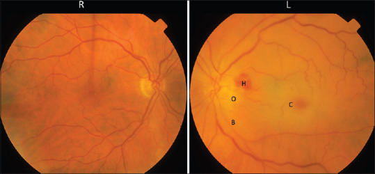
Fundus photograph showing a left acute CRAO. The color on the left is paler compared to the right retina due to retinal edema. There is disc edema (O) with peripapillary hemorrhage (H). The macula appeared erythematous called cherry-red spot (C) due to preserved choroidal circulation underlying the fovea. The retinal vessels are thin and attenuated with a boxcarring appearance (B). CRAO: Central retinal artery occlusion