
Figure 5.
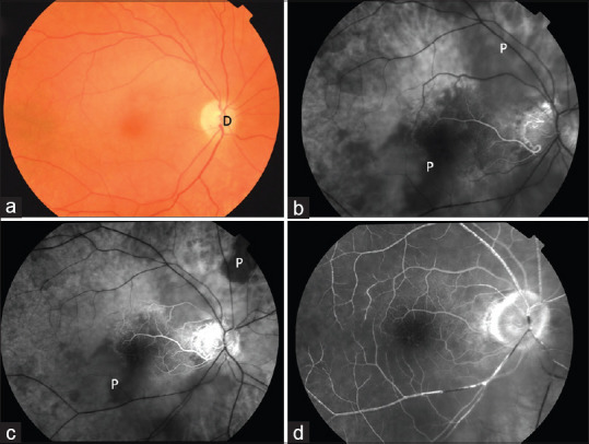
Color fundus photograph (a) of a patient with right arteritic CRAO. (b-d) FFA at 28 s, 45 s, and 1 min 28 s showing choroidal filling defect (P). CRAO: Central retinal artery occlusion
Official websites use .gov
A
.gov website belongs to an official
government organization in the United States.
Secure .gov websites use HTTPS
A lock (
) or https:// means you've safely
connected to the .gov website. Share sensitive
information only on official, secure websites.
Color fundus photograph (a) of a patient with right arteritic CRAO. (b-d) FFA at 28 s, 45 s, and 1 min 28 s showing choroidal filling defect (P). CRAO: Central retinal artery occlusion