
Figure 6.
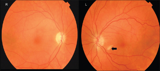
Color fundus photograph in a patient presenting with acute loss of vision due to a right CRAO. The left eye is asymptomatic but has a cotton wool spot (arrow) in the absence of other microangiopathies suggesting there is retinal nerve fiber layer ischemia. CRAO: Central retinal artery occlusion