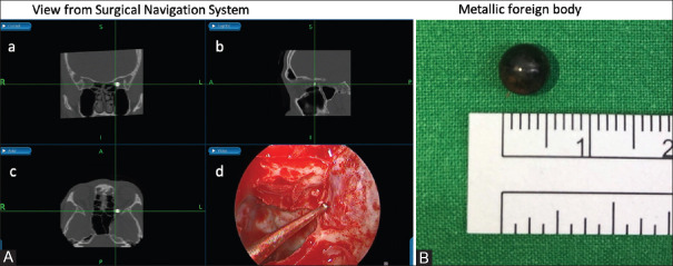
Figure 2.
Image-guidance navigation system reconstruction computed tomography image in (A-a) coronal, (A-b) sagittal, and (A-c) axial views revealed the orbital apex metallic foreign body in all three planes, and (A-d) real-time surgical view at the orbital apex. The rounded bullet was shown at the orbital apex after a small area of the lamina papyracea was removed and the periorbita incision was made; the bullet was delivered by a surgical instrument penetrated near the orbital apex. (B) The metallic foreign body was 6.0 mm in diameter

