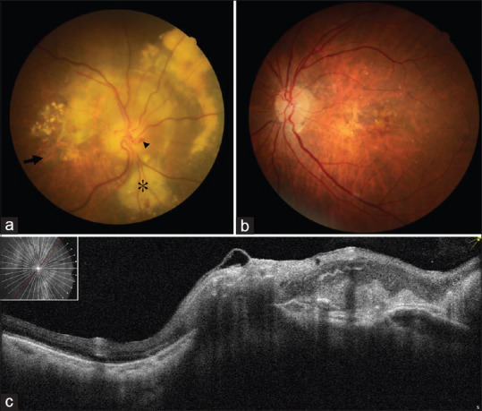
Figure 1.

(a) Fundus photograph of the right eye revealed centrally involved geographic atrophy (arrow), retinal-choroidal venous collateral (arrow head), disc edema, subretinal hemorrhage, and peripapillary exudation (asterisk). (b) Fundus photograph of the left eye revealed multiple drusen. The optic disc was normal. (c) Optical coherence tomography centered on the right eye's optic disc revealed peripapillary retina thickening and a hyperreflective area adjacent to the optic nerve head, indicating peripapillary choroidal neovascularization
