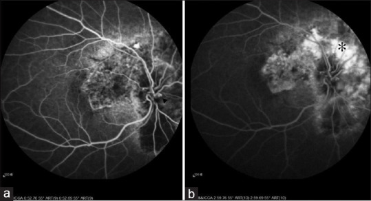
Figure 2.

(a) Fluorescein angiography demonstrated that the retinal-choroidal venous collateral (arrow head) was filled up during the arteriovenous phase in the patient's right eye. (b) Fluorescein angiography demonstrated that the peripapillary hyperfluorescence (asterisk) expanded from the superior to the nasal area of the optic disc consistent with peripapillary choroidal neovascularization
