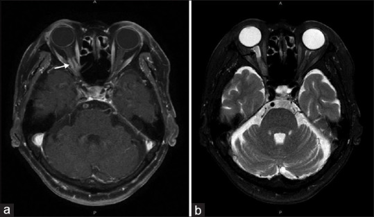
Figure 3.

(a) Aaxial T1-weighted magnetic resonance image revealed the thickening and enhancement (white arrow) of the right optic nerve sheath over the right eye's posterior orbit and optic canal, consistent with an optic nerve sheath meningioma. (b) Axial T2-weighted image revealed the increased hyperintensity (black arrow) anterior to the tumor in the right eye, indicating the accumulation of cerebrospinal fluid and distention of the perioptic subarachnoid space
