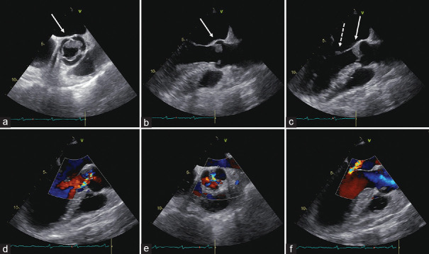
Figure 1.
Transesophageal echocardiogram. Midesophageal views at 49° (a) and 118° (b, systole; c, diastole) showing a low-isoechoic mass attached to the midportion and to free margins of the aortic cusps (white solid arrows). Aortic masses appeared low-isoechoic, with smooth margins, and homogeneous internal structure. The mitral vegetation on the anterior leaflet is indicated by the white dashed arrow (c). Midesophageal views at 134° (d) and 38° (e) showing eccentric moderate aortic regurgitation. Midesophageal views at 134° (f) moderate mitral regurgitation (central jet)

