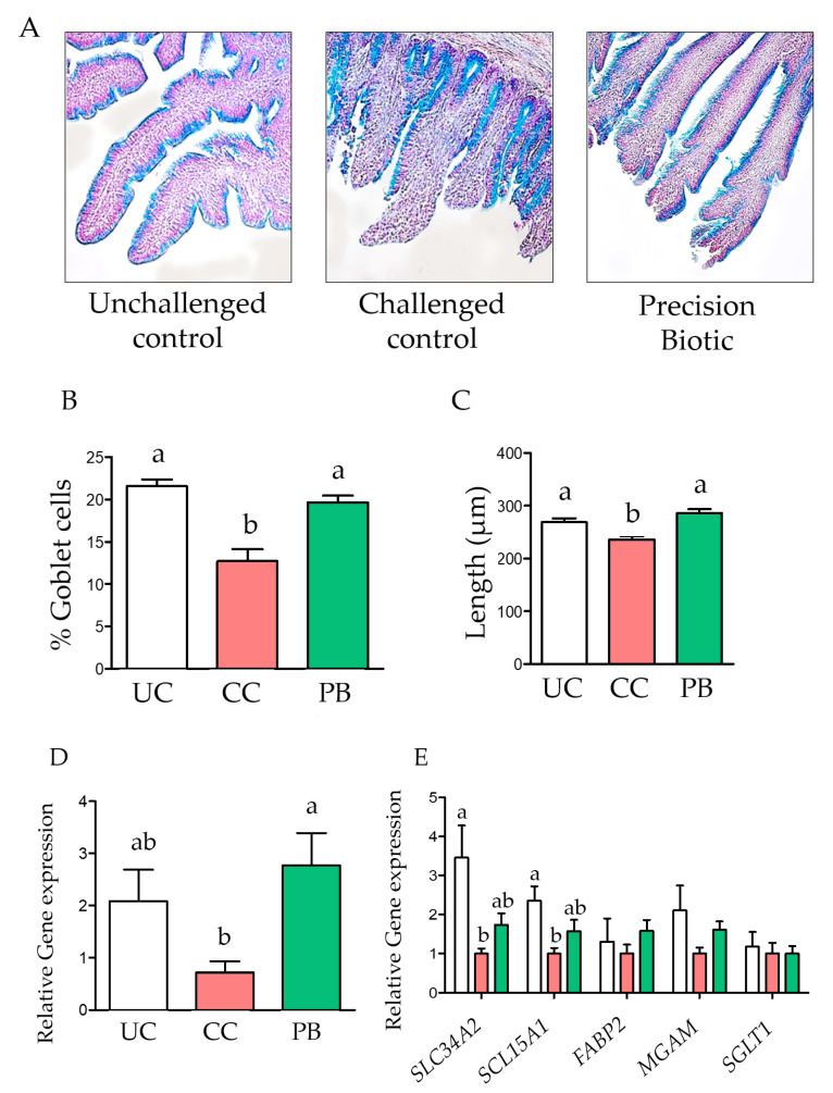
Figure 1.
A precision biotic decreased damage in villi caused by an intestinal inflammation model at 21 days of age. (A) Representative histology slides of ileal mucosa (20× magnification). (B) Percentage of goblet cells, stained with Alcian Blue. (C) Mean villi length. (D) Relative gene expression of the sodium coupled monocarboxylate transporter 1 (SLC5A8) in ileal mucosa. (E) Relative gene expression of the phosphorus transporter NaP IIb (SLC34A2), the peptide transported PepT1 (SCL15A1), the fatty acid binding protein 2 (FABP2), maltase (MAGM), and the Na+-D-glucose cotransporter (SGLT1) in ileal mucosa. One-way ANOVA with Tukey post-hoc test has been performed on all results; statistically significant differences (p < 0.05) are indicated by different letters on the bars. UC: unchallenged control; CC: challenged control; PB: precision biotic.

