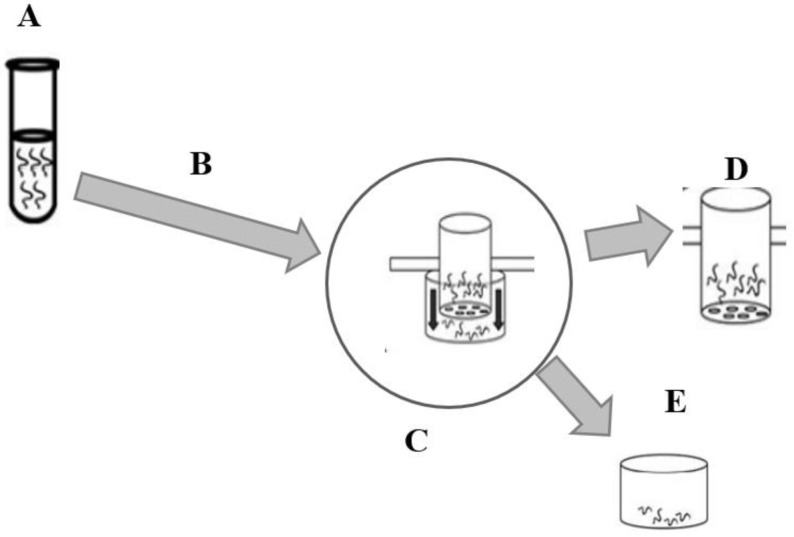
Figure 2.
Larval migration inhibition assay (LMIA) illustration. (A) Incubation of infective larvae (L3) with controls and extracts at different concentrations for 3 h (25 °C). (B) Successive washing (3 times) of incubated larvae by centrifugation (67× g) with phosphate buffered saline (PBS). (C) Transfer of washed larvae to inserts (20 µm diameter) for migration. (D) After 3 h, the larvae in the inserts were discarded. (E) Larvae count in the conical tube at the bottom of the inserts. Source: adapted from Zarate Rendon [32].

