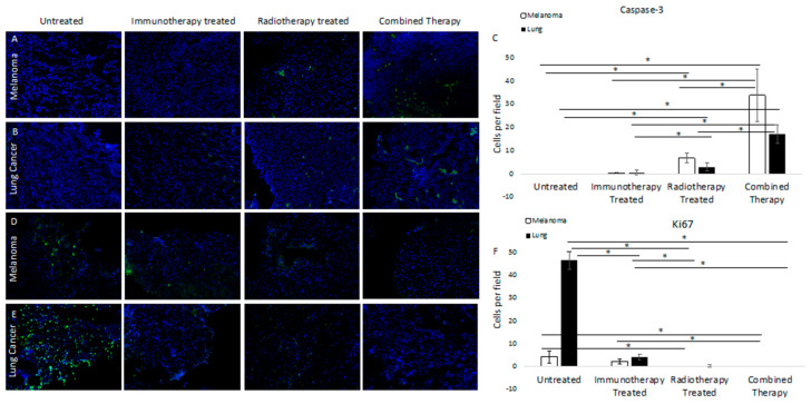
Figure 2.
Immunofluorescence staining of untreated and treated tumors for caspase-3 in melanoma (A) and lung cancer (B) demonstrated increased staining in samples treated with either immunotherapy or FAP-targeted radiotherapy, with markedly increased caspase-3 activity in tumors treated with both. (C) Quantifies positive cells per field and demonstrates significantly increased caspase-3 staining for the combined therapy group compared to untreated, immunotherapy, or radiotherapy treated samples (p = 0.001, 0.003, and 0.001 for melanoma and p = 0.0002, 0.0007, and 0.002 for lung cancer, respectively). Conversely, staining for Ki67 in melanoma (D) and lung cancer (E) demonstrated significant differences in staining of untreated samples, with decreased staining in samples treated with either therapy and essentially no Ki67 staining in tumors treated with dual therapy. (F) demonstrates quantification of positive cells per field and significantly decreased staining for the combined therapy group compared with untreated and immunotherapy-treated samples (p = 0.02, 0.003, respectively). For lung cancer, there were significant differences in Ki67 staining in the combined therapy group compared with untreated and immunotherapy-treated samples (p = 0.0000007 and 0.0005, respectively). Significant differences are marked with a line with an asterisk.

