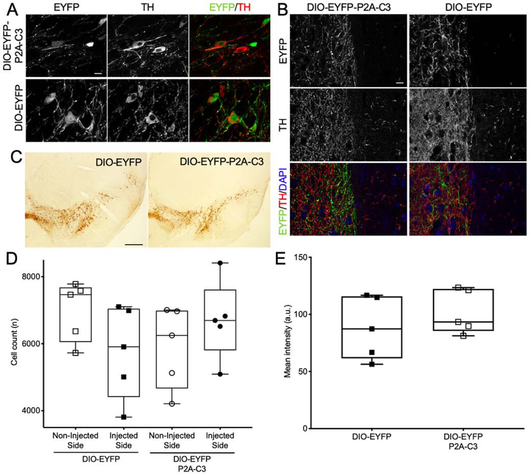
Figure 3.
No Significant Decrease in Nigrostriatal TH-Positive Neurons or TH Intensity. (3A) Confocal fluorescent micrographs demonstrating strong TH expression in dopaminergic neurons, which co-localized well with both EYFP-P2A-C3 and EYFP expression, demonstrating cell-specific expression of the floxed vectors in dopaminergic neurons of the SNc. (3B) Micrographs demonstrating expression of both C3 and TH in the striatum, indicating nigrostriatal expression of C3. (3C) TH-DAB-stained SNc sections of mice injected with either DIO-EYFP or DIO-EYFP-C3. (3D) Plot demonstrating no significant difference in mean cell counts compared between the NIS and IS in either the DIO-EYFP (n=5) or DIO-EYFP-2A-C3 (n=5) group. (3E) Plot demonstrating no significant difference in mean TH intensity between the two groups as well. Statistics: t-test. Scale bars: A) 10 μm; B) 0.5 mm; C) 10 μm.

