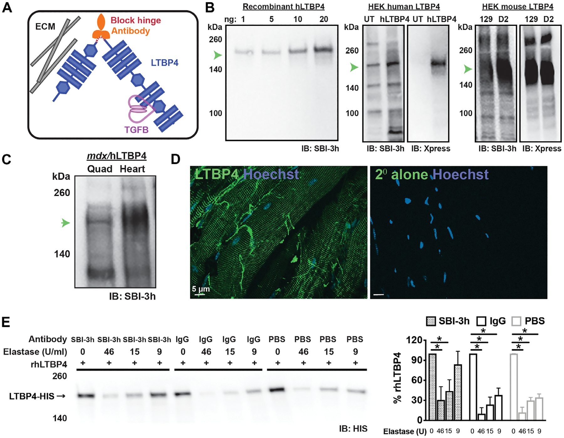
Fig. 1. Targeting the LTBP4 hinge region using a monoclonal antibody.

(A) The hinge region of LTBP4 is cleaved by serum proteases. After cleavage, latent TGFβ is released where it can be further activated; the anti-LTBP4 antibody protects LTBP4 from cleavage. ECM, extracellular matrix. (B) SBI-3h demonstrated high affinity and specificity to recombinant human LTBP4 protein and to human LTBP4 overepxressed in HEK cells. The overexpressed LTBP4 was also detected by its Xpress epitope tag. Endogenously expressed LTBP4 was also present in HEK cells [detected in the (UT, untransfected) lane]. SBI-3h also recognized the two different forms of mouse LTBP4 protein encoded by the 129T2/SvEmsJ (129) and DBA2/J (D2) substrains. IB, immunoblotting. (C) SBI-3h recognized LTBP4 protein found in lysates generated from mdx/hLTBP4 mouse muscle (quadriceps) and heart. (D) Immunofluorescence microscopic imaging of unfixed isolated myofibers showed SBI-3h (green) localized in a striated pattern. Staining of nonfixed fibers reflects LTBP4 on the external cell surface in a costameric pattern. Hoechst (blue) marks nuclei. Scale bar, 5 μm. (E) Recombinant human LTBP4 (rhLTBP4) was incubated with the high-affinity anti-LTBP4 antibody (SBI-3h), IgG control antibody, or PBS and then subjected to elastase exposure with varying concentrations of elastase. Incubation with anti-LTBP4 antibody protected against LTBP4 proteolysis seen as an increased percentage of full-length LTBP4 at 9 U of elastase. *P < 0.05 by one-way ANOVA (n = 4).
