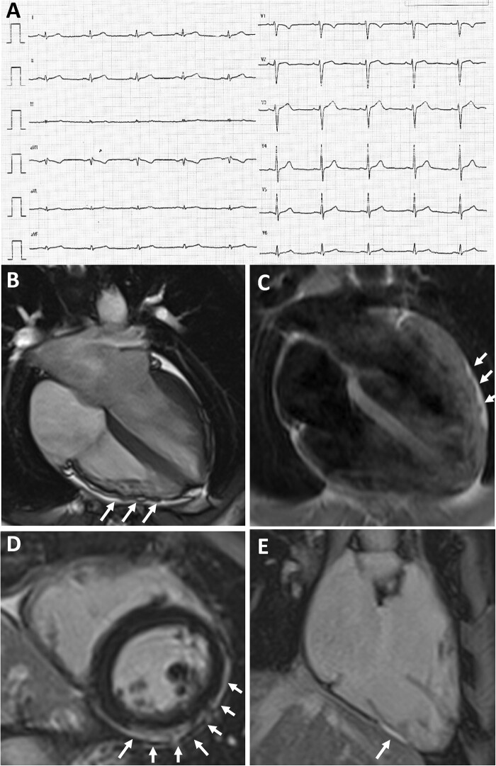
Figure 2.
Representative case of an elite athlete with low QRS-voltages underlying biventricular arrhythmogenic cardiomyopathy. A 28-year-old elite athlete engaged in a skill discipline with no family history of heart disease and no symptoms showed low QRS-voltages in the limb leads (A). Exercise testing showed premature ventricular beats with a right bundle branch block/superior axis configuration, isolated and in couples. Echocardiography was normal. CMR, cine sequences, showed hypokinesis of the mid-apical lateral LV wall and multiple small bulgings (white arrows) of the right ventricular free wall (B). On T1-weighted sequences, epicardial fatty infiltration involving the lateral and inferior LV wall (white arrows, C) and the RV free wall was observed. On post-contrast sequences, subepicardial stria of late gadolinium enhancement involving the antero-lateral, infero-lateral and inferior LV wall (white arrows on D) and the RV was detected (on inferior RV wall, white arrows on E). Genetic testing was negative. Based on RV dilation plus wall motion abnormalities on CMR and non-sustained ventricular tachycardia on 24-hour Holter monitoring a diagnosis of arrhythmogenic cardiomyopathy was made, that was ‘borderline’ according to the 2010 Task Force Criteria and ‘definite’ according to the 2020 International criteria (Padua criteria) that also incorporate tissue characterization findings by CMR.12,13 CMR, cardiac magnetic resonance; LV, left ventricular; RV, right ventricular.

