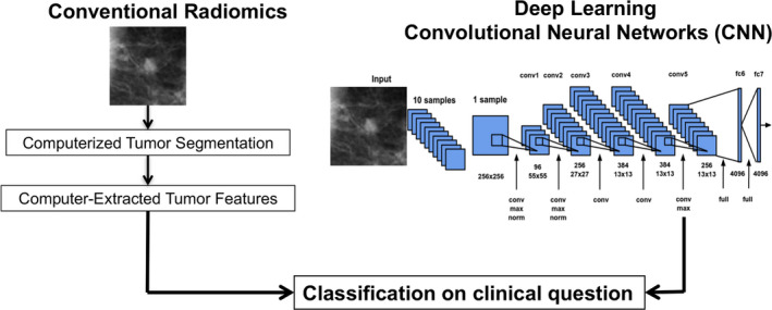
Figure 2.

Two general classification approaches to obtaining quantitative data for radiomics; (left) conventional manual or semi‐automated lesion segmentation followed by computer‐extracted features. and (right) fully automated deep convolutional neural networks. Here, the clinical task is in distinguishing between malignant and benign breast lesions. Both methods can take advantage of machine learning techniques and the need to train from annotated known cases. From Ref. 8.
