Figure 3.
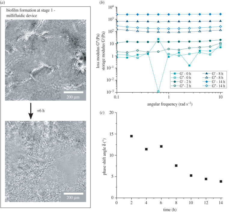
Development of B. subtilis biofilm grown in flow (LBGM medium at 30°C with flow speed v = 346 μm s−1) over time. (a) Growth during stage 1: phase contrast microscopy images of the network formation of B. subtilis biofilm during stage 1 in the millifluidic device, which holds the detachable measurement geometry (depicted in figure 2a). Images were taken at the midplane, 100 μm away from the vertical and horizontal channel surfaces. In phase contrast, the cells appear black while the biofilm matrix appears white. (b) Frequency sweeps of biofilms grown with constant nutrient flow during the growth phase on the rheometer, after 2 days of growth in the separate millifluidic device. (c) Phase shift angle taken at ω = 1 rad s−1 from the frequency sweeps, which were performed every 2 h during growth.

