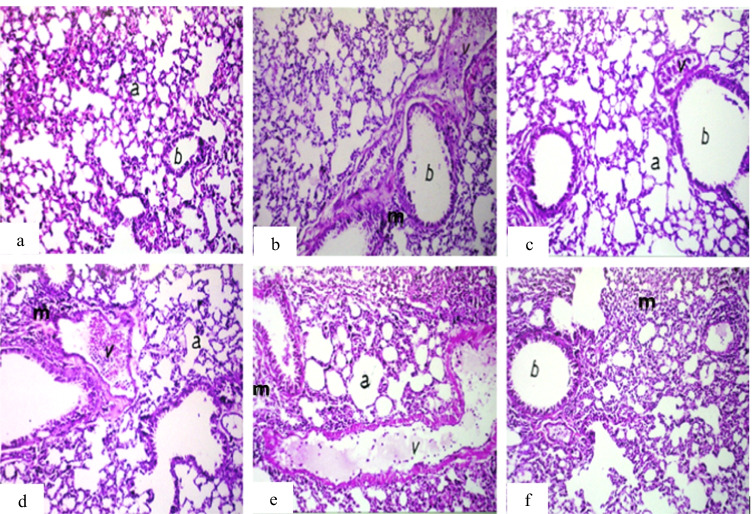
Fig. 8.
Photomicrograph of lung sections of mice. a Group 1 (control) showing normal histological structure of the alveoli (a) and bronchioles (b). b Group 2 (IP-MNPs) showing peribronchiolar inflammatory cell infiltration (m). c Group 3 (IM-MNPs) showing emphysema in air alveoli (a) with congestion in peribronchiolar blood vessels (v). d Group 4 (ESC) showing dilatation of the blood vessels (v) with perivascular inflammatory cell infiltration (m) and emphysema in the air alveoli (a). e Group 5 (ESC + IP-MNPs) showing dilatation and congestion of the blood vessels (v) with emphysema in the air alveoli (a) and inflammatory cell infiltration in between (m). f Group 6 (ESC + IT-MNPs) showing diffuse inflammatory cell infiltration in the air alveoli (m). H&E, 40 ×

