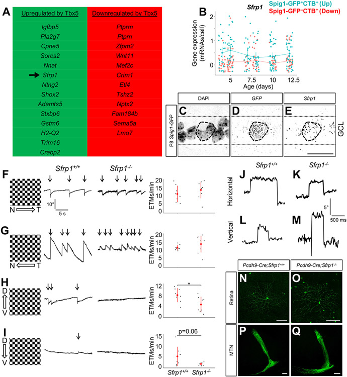
Figure 4. Sfrp1 is a potential downstream effector of Tbx5.
(A) Cross-referencing bulk RNAseq data from Tbx5+/+ and Tbx5−/− cardiac tissue40 with our oDSGC single-cell RNAseq data yielded a list of genes potentially upregulated or downregulated by Tbx5 (rank-ordered by q-value). (B) Sfrp1 was expressed at slightly higher levels in up-oDSGCs compared to down-oDSGCs between P4 and P12. Data presented as mean ± 95% confidence intervals. (C-E) RNAscope in situ hybridization in P8 Spig1-GFP retinas shows Sfrp1 expression in GFP+ up-oDSGCs within the ganglion cell layer (GCL). (F-I) Optokinetic reflex (OKR) measurements in adult Sfrp1+/+ and Sfrpt−/− mice showed comparable performance in response to nasotemporal (F) and temporonasal (G) motion. Sfrpt−/− mice showed impaired OKR performance in response to ventrodorsal motion (H) and a trend to statistically significant impairment in response to dorsoventral motion (I). Data presented as mean ± SD. (J-M) Voluntary horizontal (J-K) and vertical (L-M) saccades were intact in Sfrp1−/− mice. (N-Q) Adult up-oDSGCs, labeled with intraocular AAV2 FLEX-GFP in Pcdh9-Cre;Sfrp1+/+ and Pcdh9-Cre;Sfrp1−/− mice, appeared grossly normal with regards to dendrite morphology (N-O) and axon projections to the MTN (P-Q). Scale bars, 25 um in (C-E); 100 um in (N-Q). *p < 0.05. ETM, eye-tracking movement. See also Figure S4.

