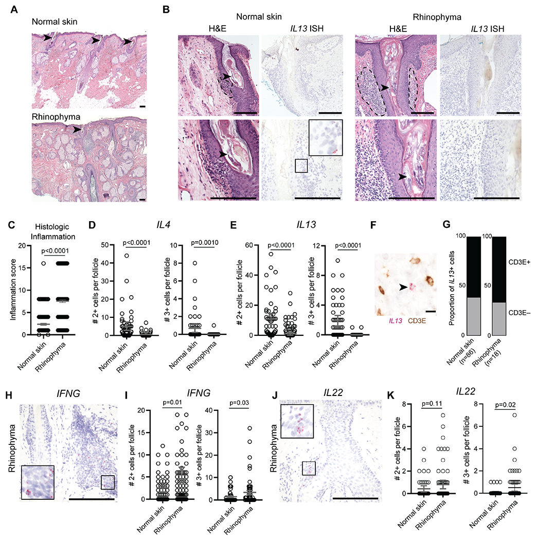
Figure 2. Type 2 cytokines expression is associated with healthy Demodex commensalism in humans.

(A) Low power H&E stained sections of normal skin (top) and rhinophyma (bottom). (B) H&E stained sections (left panels) and corresponding IL13 mRNA in situ hybridization (ISH, right panels) in hair follicles infected with Demodex in representative cases of normal skin and rhinophyma. Arrowheads highlight Demodex infection of the hair follicle. Dashed lines highlight perifollicular inflammation. Scale bars, 250 μm. (C to E) Scoring of hair follicle inflammation (C) and quantification of IL4 (D) and IL13 (E) expression by ISH. (F and G) Representative co-stained section for IL13 by ISH and CD3ε by immunohistochemistry (F) and quantification of proportion of CD3+ or CD3− IL13-expressing cells (G). Arrowhead highlights IL13+CD3− cell. Scale bar, 10 μm. (H and I). Representative IFNG mRNA ISH (H) and quantification of IFNG expression (I) in hair follicles infected with Demodex. (J and K). Representative IL22 mRNA ISH (J) and quantification of IL22 expression (K) in hair follicles infected with Demodex. Scale bars, 250 μm. Statistical significance shown by * P < 0.05, ** P < 0.01, *** P < 0.001, **** P < 0.0001 by two-tailed Student’s t test.
