FIGURE 1.
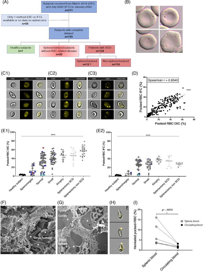
Proportion of Pocked RBC in the circulation: Inter-method consistency, correlation with spleen size, progressive pathological processes, RBC filtration, and pitting in SCD spleens. (A) Flow chart of subjects and patients. Quantification of pocked-RBC by IFC was performed in 7 of 12 splenectomized patients with SCD (A). (B) Typical aspects of single- and multiple-pocked-RBC by DIC. (C) Typical aspects of spotless RBC (C1), single- (C2) and multiple-spotted RBC (C3) by IFC. Scale bars = 5 μM. (D) Linear regression relationship and nonparametric Pearson correlation between % pocked-RBC by DIC and IFC in 221 blood samples (Y = 0.6277*X + 4.777), (****: p < .0001). In 63 instances, patients were sampled pre- and post-transfusion (126 samples), 15 patients were sampled twice and 1 patient was sampled 4 times during follow-up. (E) Proportion of pocked-RBC in peripheral blood samples collected before transfusion, assessed by DIC (E1) and IFC (E2) in different subgroups of adult patients with SCD and healthy individuals defined by spleen size. Each dot represents a single measure or mean of measures when the patient had been sampled multiple times. Genotypes and status regarding splenectomy are differentiated as follows: HbSS (gray dots, gray crosses for SCD splenectomized subjects), HbS/beta-zero (red dots), HbSC (blue dots), HbS/beta-plus (cyan dots), and splenectomized subjects without underlying RBC disease (black crosses). Kruskal-Wallis and Dunn’s multiple comparison tests were used for both the whole cohort and HbSS separately (****: p < .0001). Black bar represents the statistical analysis including all genotypes. (F) Scanning electron microscopy of spleen sample from an SCD child showing packed RBC, many of which are sickled (star) and a typical sinus structure with the cordal side on the left and the luminal side on the right, elongated endothelial cells (EC), basal helicoid fibers (horizontal arrowheads), both delimiting an inter-endothelial slit (vertical arrowheads) into which an RBC is engaged (only its cordal part is visible, oblique arrow) (F). (G) Transmission electron microscopy shows a pocked-RBC crossing an inter-endothelial slit, the vacuole-containing part of the RBC lying upstream from the slit (oblique arrow). Sickled RBC are visible in the sinus lumen and cords (stars). (H) Typical images of “herniated” pocked-RBC, and their proportions in circulating blood and spleen blood from 6 children with SCD (I). Wilcoxon matched-pairs signed rank test was used (*: p < .0332)

