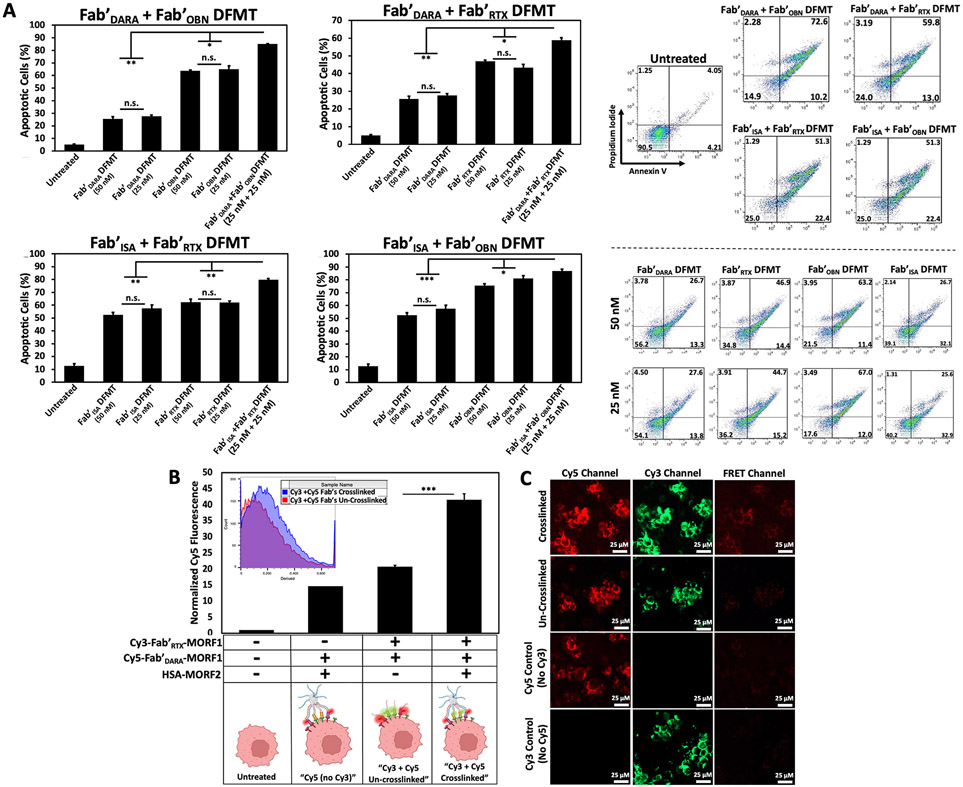
Fig. 2.
(A) Heteroreceptor DFMT-induced apoptosis on CD20+ / CD38+ Raji cells. Cell viability was measured by Annexin V and Propidium Iodide staining and analyzed by flow cytometry. Flow cytometry cell population distribution for all four heteroreceptor combinations are shown, along with representative cell population distributions for individual DFMT treatments at [MORF1] = 25 nM and 50 nM. (B) Flow cytometry output of FRET-induced excitation of Cy5-Fab'DARA-MORF1 upon excitation of Cy3-Fab'RTX-MORF1 on the cell surface. FRET emission was quantified as the amount of fluorescence observed when exciting cells with a 488 nm excitation with 530 / 30 nm band-pass filter and detecting emission 670 / 30 nm. (C) Confocal microscopy images of Cy3-Fab'RTX-MORF1 and Cy5-Fab'DARA-MORF1 treated Raji cells with or without crosslink-inducing macromolecule treatment. FRET-induced emission of Cy5 can be observed in the crosslinked treatment group, but no emission detectable in the un-crosslinked treatment group. Unlabeled Fab'RTX-MORF1 was used for “No Cy3“ control and unlabeled Fab'DARA-MORF1 was used for “No Cy5“ control. Both controls were crosslinked. All experiments were performed in triplicate. *** p < 0.001, ** p < 0.01, *p < 0.05, n.s. not significant by One-Way ANOVA and Tukey test.

