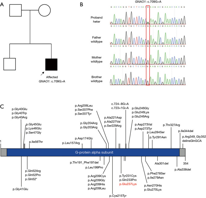
Figure 2.
Family pedigree—Sanger sequencing. (A) The pedigree of our patient. The affected proband is highlighted by the square and arrow. (B) The variant in the GNAO1 gene was confirmed by Sanger sequencing in the trio family. The mutated site is shown in the red box. (C) This is the schematic of the variants reported thus far. A total of 50 amino acid variations and the domain G-protein alpha subunit are shown. The variant in our patient is highlighted with red text.

