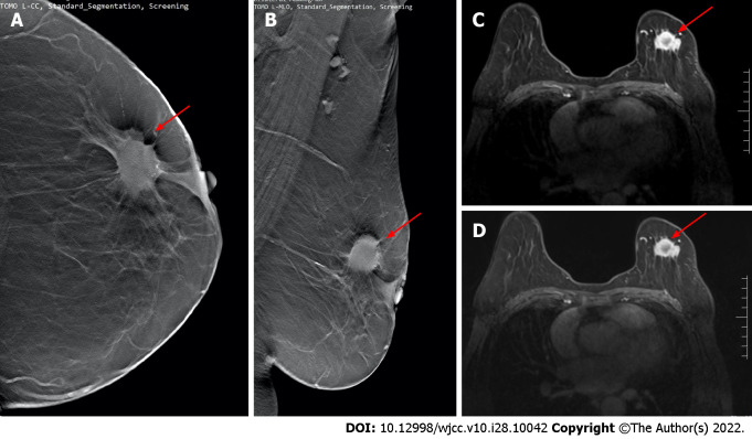
Figure 2.
Digital breast tomosynthesis and magnetic resonance imaging examination. A and B: Images of digital breast tomosynthesis examination. Irregular lumps in the upper quadrant of the left breast and burr signs can be seen on the periphery (red arrows); C and D: Magnetic resonance imaging examination images of the same patient. The irregular lumps on the left breast show obvious postoperative disease (red arrows). It was confirmed by physiology as stage II invasive ductal carcinoma.

