. 2022 Aug 3;28(20):4536–4550. doi: 10.1158/1078-0432.CCR-22-0568

Figure 3.

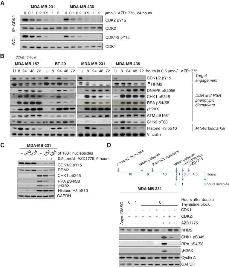
Figure 3. WEE1 inhibition results in activation of CDK2 and subsequent DNA damage and replication stress response in breast cancer cell lines. A, Immunoblot analysis showing CDK2 and CDK1 activation 24 hours after treatment with different doses of AZD1775 in MDA-MB-231 and MDA-MB436 cells. To assess CDK2 tyrosine phosphorylation (pY15) level, total CDK2 was first immunoprecipitated (IP) and the bound fraction were eluted and analyzed. WCL, whole cell lysate; IP, immunoprecipitation. B, Immunoblot analysis of TNBC cell lines treated with DMSO (U) or AZD1775 0.5 μmol/L during different time periods. Biomarkers of target engagement, DNA damage response (DDR) and replication stress response (RSR) or mitosis were analyzed. C, Immunoblot analysis of target engagement and DDR biomarker in MDA-MB-231 cells treated with DMSO (−) or 0.5 μmol/L AZD1775 for 6 hours in the presence (+) or absence (−) of diluted EmbryoMax nucleoside solution. D, Immunoblot analysis of MDA-MB-231 cells from S-phase culture synchronized by double thymidine block treated with DMSO (−) or AZD1775 0.5 μmol/L in the presence (+) or absence (−) of RO-3306 (CDK1 inhibitor, CDK1i) or CVT-313 (CDK2 inhibitor, CDK2i). Protein samples were collected at indicated time points and the indicated biomarkers were analyzed.

WEE1 inhibition results in activation of CDK2 and subsequent DNA damage and replication stress response in breast cancer cell lines. A, Immunoblot analysis showing CDK2 and CDK1 activation 24 hours after treatment with different doses of AZD1775 in MDA-MB-231 and MDA-MB436 cells. To assess CDK2 tyrosine phosphorylation (pY15) level, total CDK2 was first immunoprecipitated (IP) and the bound fraction were eluted and analyzed. IP, immunoprecipitation; WCL, whole cell lysate. B, Immunoblot analysis of TNBC cell lines treated with DMSO (U) or AZD1775 0.5 μmol/L during different time periods. Biomarkers of target engagement, DNA damage response (DDR) and replication stress response (RSR) or mitosis were analyzed. C, Immunoblot analysis of target engagement and DDR biomarker in MDA-MB-231 cells treated with DMSO (−) or 0.5 μmol/L AZD1775 for 6 hours in the presence (+) or absence (−) of diluted EmbryoMax nucleoside solution. D, Immunoblot analysis of MDA-MB-231 cells from S-phase culture synchronized by double thymidine block treated with DMSO (−) or AZD1775 0.5 μmol/L in the presence (+) or absence (−) of RO-3306 (CDK1 inhibitor, CDK1i) or CVT-313 (CDK2 inhibitor, CDK2i). Protein samples were collected at indicated time points and the indicated biomarkers were analyzed.