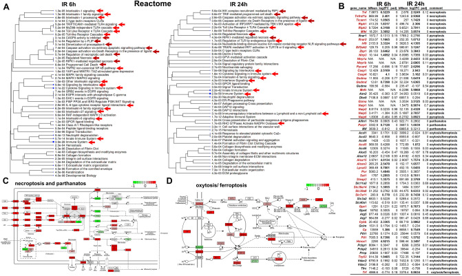
Figure 4.
Multiple types of programmed necrosis are active simultaneously in the IR retina. (A) The results of the pathway analysis revealed that many genes with increased expression in the ischemic retina are involved in programmed (regulated) necrosis (red arrows indicate pro-inflammatory signaling cascades, dark red arrows indicate signaling cascades involved in programmed necrosis). (B) The expression of many genes regulating necroptosis, pyroptosis, oxytosis/ferroptosis, and parthanatos is significantly increased in the ischemic retina 6 and 24 h after reperfusion. We highlighted in dark red those genes whose expression was significantly increased (two-fold or more) in IR retinas. (C, D) The representative KEGG pathway maps show increased expression of necroptosis, oxytosis/ferroptosis, and parthanatos genes in the IR retina. The red box corresponds to increased gene expression in the ischemic retina, the green box corresponds to reduced gene expression in the ischemic retina.

