Figure 6.
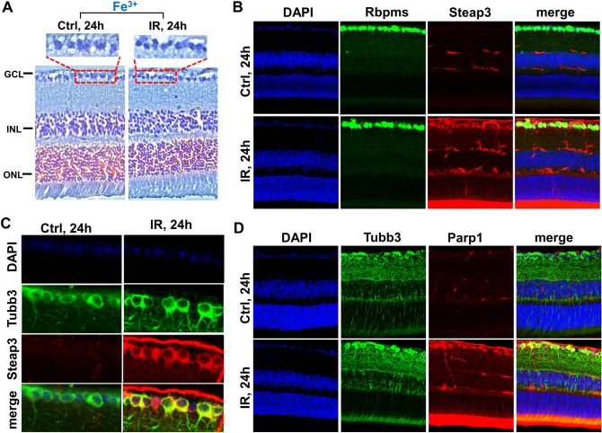
The expression of genes involved in oxytosis/ferroptosis, and parthanatos is significantly increased in ischemic RGCs. (A) Prussian blue stain indicates a high content of iron in neurons of ischemic and normotensive retinas. Prussian blue is specific for ferric iron (Fe3+). (B, C) Immunohistochemistry (IHC) analysis reveals that Steap3, the key enzyme generating ferrous (Fe2+) iron from ferric (Fe3+) iron, is highly expressed in the ischemic retina. Rbpms and Tubb3 are RGC markers. (D) IR results in high expression of Parp1 in RGCs.

