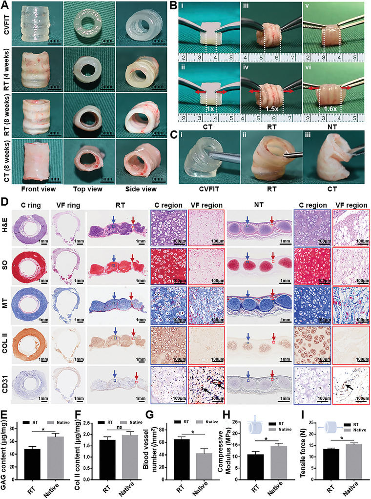
Figure 5.

In vivo subcutaneous implantation of bioprinted CVFIT in nude mice. A) Gross view of the bioprinted CVFIT, regenerated trachea, and unitary cartilage tube after 4 or 8 weeks of implantation. B) Mechanical function evaluations of longitudinal tensile and C) lateral bending tests. CT: cartilage tube; RT: regenerated trachea; NT: native trachea. D) Histological examinations of H&E, safranin‐O (SO), Masson's trichrome (MT), type II collagen (COL II), and CD31 staining of the regenerated trachea in the transverse and longitudinal sections at 8 weeks and of the native counterpart. Blue arrows represent cartilage regions; red arrows represent vascularized fibrous tissue regions; black arrows represent blood vessels. Quantitative analyses of the E) GAG contents, F) COL II contents, G) blood vessel numbers, H) Compressive moduli, and I) tensile forces of the regenerated and native trachea (n = 4, *p < 0.05, ns: no significance).
