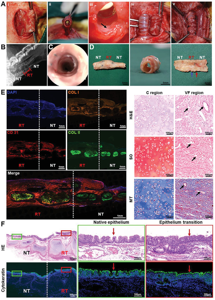
Figure 6.

Segmental trachea reconstruction surgery and therapeutic outcome. A‐i,ii) Photographs of regenerated trachea with the muscle pedicles, iii) well‐vascularized inner wall of regenerated trachea, iv) exposed regenerated and native trachea, and v) end‐to‐end anastomosis of regenerated trachea with cut ends of the native trachea. Black arrows represent blood vessels. B) X‐ray and C) tracheoscopy images of the in situ repaired trachea segment at 8 weeks postsurgery. D) Gross view of the reconstructed trachea segment at 8 weeks postsurgery. Blue arrows represent the regenerated cartilage ring; red arrows represent regenerated vascularized fibrous ring. E) Histological staining of H&E, Safranin‐O (SO), Masson's trichrome (MT), and immunofluorescence staining of type II collagen (COL II, green), type I collagen (COL I, orange), CD31 (red), and cell nuclei (DAPI, blue). Black arrows represent blood vessels. F) H&E staining and immunofluorescence staining of cytokeratin (green) and cell nuclei (DAPI, blue) showing the regeneration of tracheal epithelium. Red arrows represent mucosaepithelium. RT: regenerated trachea; NT: native trachea.
