FIGURE 3.
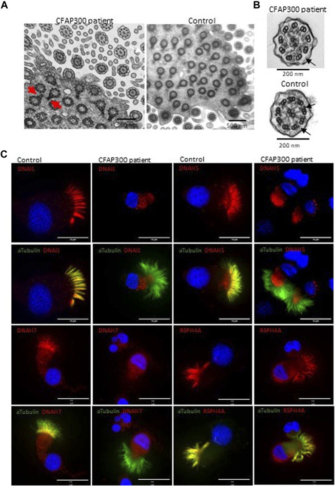
Localization of axonemal proteins in airway cilia of a control and PCD patient (patient II-1) with CFAP300 mutations. (A). High number of cilia cross sections were present in the nasal brushing sample of a PCD patient with CFAP300 c.198_200delinsCC mutations, but the orientation of the basal foot (red arrows) was disorganized compared to control. (B). The main finding in TEM of cilia cross sections in a patient with the CFAP300 variant was lack of dynein arms (black arrows). (C). ODA (DNAH5 and DNAI1) and IDA (DNAH7) proteins were missing from cilia in the patient airway epithelial cilia, while strong staining is present in the control sample. Radial spoke protein RSPH4A is present in the patient cilia. αTubulin was used as a marker for cilia and Dapi as a nuclear stain. Scale bar is 10 µm.

