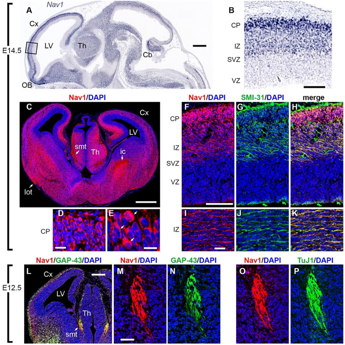
FIGURE 1:
Nav1 is localized in neuronal cell bodies and growing axons in developing mouse brain. (A, B) Nav1 mRNA, detected by in situ hybridization (ISH) on E14.5 (Genepaint; set ID EH1164), was widely expressed in differentiating neurons but not in progenitor cells around the ventricles. In the cerebral cortex (boxed area in A, shown at higher magnification in B), Nav1 mRNA was not detected in the progenitor-containing ventricular zone (VZ) and subventricular zone (SVZ) but was highly expressed in the intermediate zone (IZ) and cortical plate (CP), where postmitotic neurons are located (Genepaint; set ID EH1164). (C–E) Nav1 protein on E14.5 was expressed in neuronal cell bodies but was also highly enriched in axon tracts including the stria medullaris thalami (smt), lateral olfactory tract (lot), and internal capsule (ic). Higher-magnification views of the CP in D and E demonstrate Nav1 protein in many cell bodies (arrows in E). (F–K) Double immunofluorescence for Nav1 and neurofilament heavy chain (antibody SMI-31), a marker of axons, confirmed the presence of Nav1 in axons coursing through the IZ. (L–N) On E12.5, double immunofluorescence for Nav1 and GAP-43, a marker of growth cones, demonstrated extensive colocalization, notably in growing axons of the smt, shown at higher magnification in M and N. (O, P) Double immunofluorescence for Nav1 and βIII-tubulin (antibody TuJ1), a marker of neurons and their processes, confirmed the presence of Nav1 in smt axons. Plane of section: sagittal for A, B; coronal for C–P. Scale bars: A, 500 μm; B, 100 μm; C, 500 μm; D, 20 μm; E, 10 μm; F–H, 100 μm; I–K, 20 μm; L, 200 μm; M–P, 50 μm. Cx = Cortex, LV = Lateral Ventricle, Th = Thalamus, OB = Olfactory Bulb, Cb = Cerebellum. Images represent observations from three individual culture preparations.

