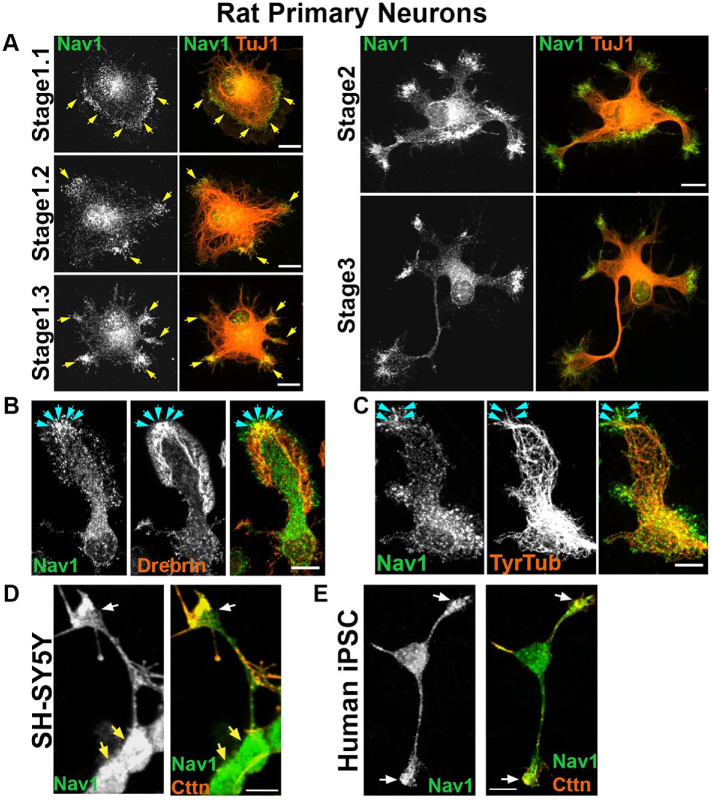
FIGURE 2:
Nav1 is expressed in areas of morphological change. (A) Primary cultures from rat hippocampus were fixed after 3 DIV and immunostained for Nav1 and neuron-specific βIII-tubulin (antibody TuJ1). Representative images of neurons at different stages of neuritogenesis are shown. In stage 1.1 neurons have a lamellipodium surrounding the cell body and Nav1 is enriched close to the membrane in this area. As the lamellipodium undergoes segmentation (stage 1.2), Nav1 remains enriched in this location. The segments coalesce into nascent growth cones as the MTs adopt a parallel organization behind them (stage 1.3). Nav1 remains similarly clustered in nascent growth cones as minor neurites become established in stage 2, and one neurite is specified as the presumptive axon in stage 3. (B) Representative image of a growth cone immunostained for Nav1 and the actin-binding protein drebrin, highlighting the transition zone of the growth cone. (C) Representative image of growth cone immunostained for Nav1 and tyrosinated tubulin shows that newly polymerized MTs are present together with Nav1. (D) Representative image of 3 d BDNF-differentiated SH-SY5Y cells showing enriched Nav1 puncta in distal ends of neurite (white arrows indicate growth cone yellow arrows indicate cell bodies). (E) Representative image of human iPSC–derived neuron showing enriched Nav1 in distal ends of neurites (white arrows indicate growth cones). All scale bars = 10 μm. Images represent observations from three individual culture preparations.

