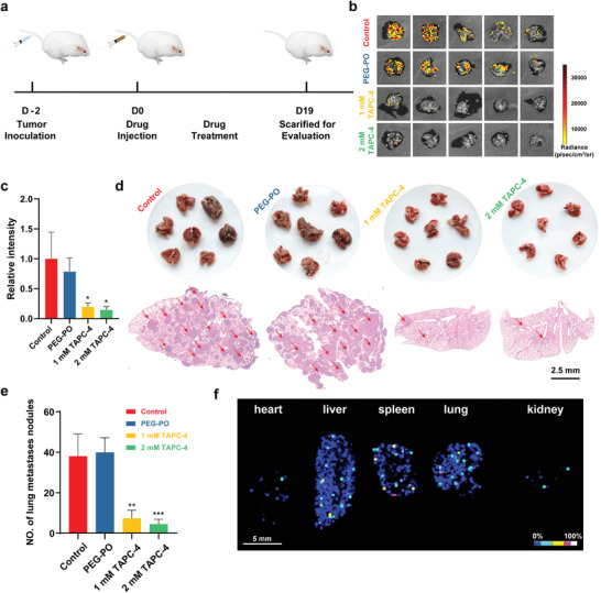
Figure 6.

TAPC‐4 inhibits lung metastasis of melanoma in vivo. a) Scheme of lung metastatic melanoma model fabrication and treatments with saline, PEG‐PO, and PEG‐PO modified TAPC‐4 at different doses (n = 7). b) The lung luminescent images and c) relative luminescent intensity of B16‐F10‐Luc cells in the lung tissues (n = 5). d) Top: Macroscopic images of lung tissues. Bottom: H&E‐stained lung slices. The scale bar is 2.5 mm. e) Quantification of lung metastatic nodules (n = 7). f) MALDI‐TOF‐MS imaging of TAPC‐4 in primary organs. Mice were treated with 1 mm TAPC‐4 for 24 h, and the characteristic MS signal of C60 at 720 m/z was collected for the imaging (Mean ± SEM; Student's t‐test, *p < 0.05, **p < 0.01, and ***p < 0.001).
