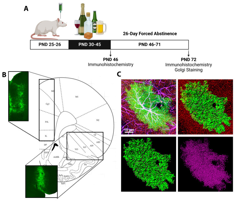
Figure 1.
Experimental Design: (A) Animals received intracranial injections of an astrocyte-specific adeno-associated virus directly into the ACC, mPFC, or OFC on PND 25–26. Beginning PND 30, animals received 5 g/kg of EtOH or water via oral gavage on an intermittent, 2 days on, 1 day off, 2 days on, 2 days off, schedule. Tissue was collected on PND 46, during peak withdrawal, or after a 26-day forced abstinence period, on PND 72. (B) Validation of Lck-GFP expression in the ACC and mPFC (upper left image) and the OFC (bottom image). (C) Representative images of AAV+ astrocyte imaging with confocal microscopy and reconstruction using Imaris. The top left image is a confocal image of an AAV+ astrocyte (green), with GFAP (magenta), co-localization of AAV+ and GFAP (white), and PSD-95 (red), scale bar, 10 µm. The top right image is a reconstructed astrocyte (green) and PSD-95 (red). The bottom left image is a reconstructed surface rendered astrocyte (green) with PSD-95 (magenta) within 0.5 µm of the astrocyte. The bottom right image is isolated PSD-95 (magenta) that is colocalized with the astrocyte of interest.

