
Figure 1.
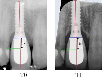
Radiograph taken at prosthesis placement (reference T0) and at the long‐term control (T1). The implant length was measured on each radiograph and compared to that given by the manufacturer for internal calibration. Reproducible landmark (green) on the adjacent teeth was selected on T0 and T1 radiographs. The projection of the T0 landmark (green) on the long axis of the implant only (red) and not the crown established the baseline distance X1. The same measurement was made on the T1 image. X2 was then subtracted to X1 to determine the amount of vertical discrepancy between T0 and T1.