Figure 7.
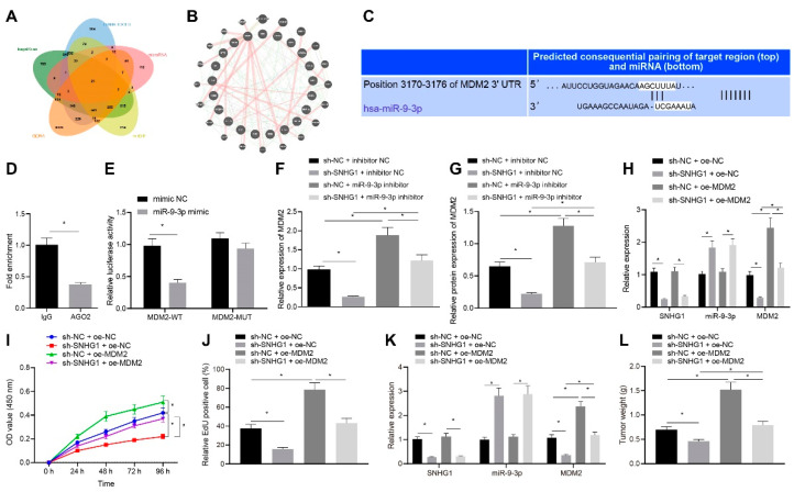
SNHG1 silencing decreases MDM2 expression via miR-9-3p to repress bladder cancer cell proliferation and tumorigenesis. (A) The downstream genes of miR-9-3p predicted by TargetScan: http://www.targetscan.org/vert_71/ (accessed on 8 August 2021), DIANA TOOLS: http://diana.imis.athena-innovation.gr/DianaTools/ (accessed on 8 August 2021), microRNA: http://www.microrna.org/microrna/home.do (accessed on 8 August 2021), and mirDIP: http://ophid.utoronto.ca/mirDIP/ (accessed on 8 August 2021). (B) The differential genes in bladder cancer obtained by GEPIA analysis of TCGA database. (C) Binding sites between miR-9-3p and the 3′UTR of MDM2 predicted by TargetScan website. (D) RIP experiment finding that AGO2 could pull down MDM2. (E) Targeting relationship between miR-9-3p and MDM2 assessed by dual-luciferase reporter gene assay. (F) The expression of MDM2 in T24 cells after silencing both SNHG1 and miR-9-3p detected by qRT-PCR. (G) The expression of MDM2 in T24 cells after silencing both SNHG1 and miR-9-3p detected by Western blot analysis. (H) The expression of SNHG1, miR-9-3p and MDM2 after overexpressing MDM2 and silencing SNHG1 in T24 cells measured by qRT-PCR. (I) CCK-8 assay of the change of T24 cell viability. (J) The changes of T24 cell proliferation detected by EdU assay. (K) qRT-PCR detection of the expression of SNHG1 and miR-9-3p in T24 cells. (L) Tumor weight. * p < 0.05. N = 10 mice in each group. The cell experiment was repeated three times. Data were presented as mean ± standard deviation and compared by one-way analysis of variance or unpaired t test.

