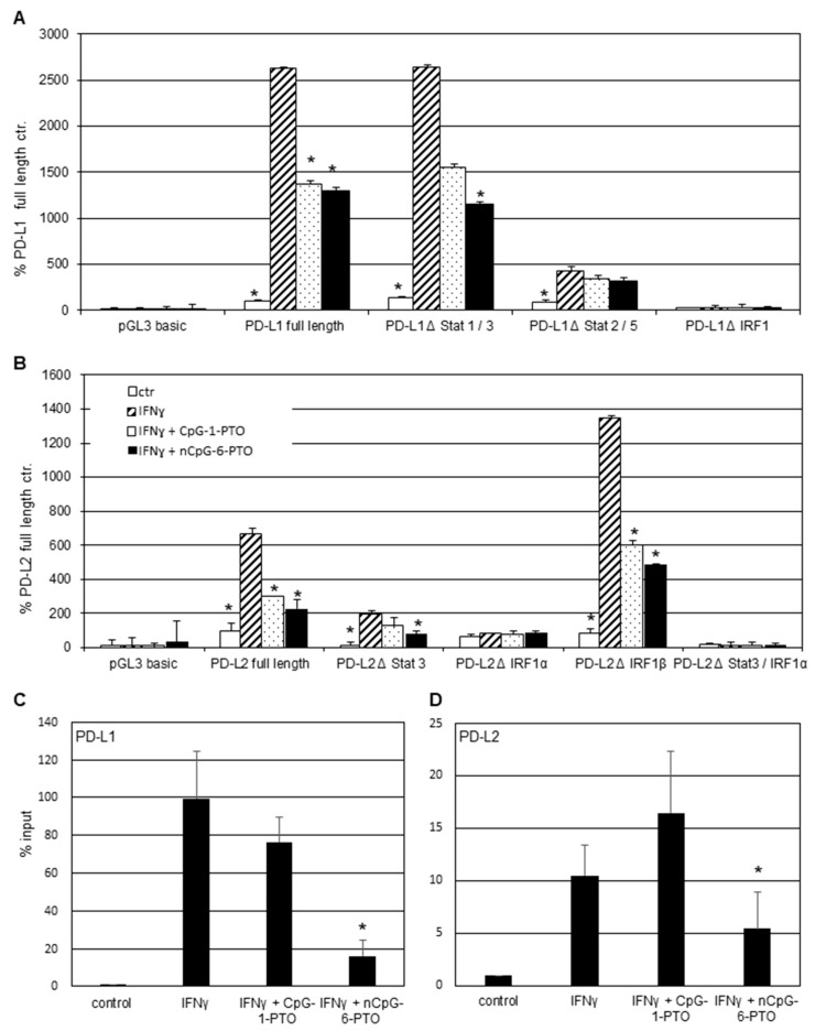
Figure 4.
Promoter function analysis. Transient luciferase reporter assays for the (A) PD-L1 and (B) PD-L2 promoter. A375 melanoma cells were transfected with PD-L1 and PD-L2 promoter constructs including deletions of the relevant transcription binding sites. After stimulation with 20 ng/mL IFNγ for 1 h, cells were treated with 4 µM CpG-1-PTO or nCpG-6-PTO for 16 h. (C) ChIP assay after pretreatment with 4 µM CpG-1-PTO or nCpG-6-PTO for 1 h and consecutive IFNγ stimulation for 6 h using the IRF1 antibody for precipitation. PCR was performed with PD-L1 or (D) PD-L2 promoter specific primers. Each column represents the mean of 3 experiments. Statistical analysis was performed in relation to controls treated with IFNγ solely; * p < 0.05.

