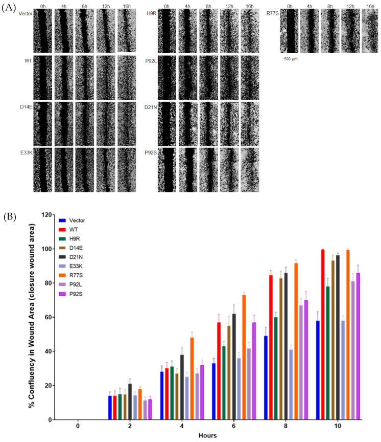
Figure 4.
Migration of stably transfected NIH3T3 cell lines in a wound healing (scratch) assay. Stable NIH3T3 cell clones were plated onto 24-well plates. Wounds in the monolayer were created with an automated tool, plates were washed to remove detached cells, and closure of the wound was followed for 24 h with hourly imaging in a BioTek Lionheart FX Automated Live Cell Imager. Images are shown in (A) and quantitation over time in (B). Results are expressed relative to WT E7 migration at 10 h. At 10 h: WT vs. Vector (p = 0.0022); WT-H9 R (p = 0.0079); WT-E33K (p = 0.0022); WT-D14E (p > 0.05); WT-P92L (p = 0.0080) WT-P92S (p = 0.0082). If we apply a highly conservative Bonferroni multiple testing correction of six tests of mutant versus WT, only vector and E33K remain significant.

