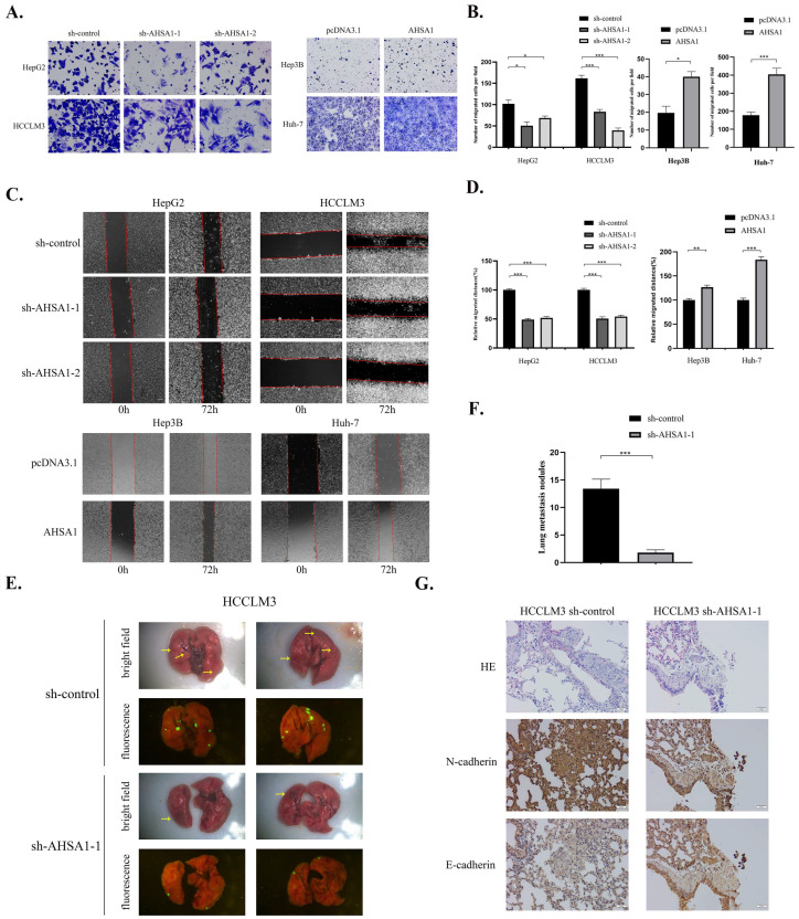
Figure 3.
AHSA1 promoted the invasion, migration, and EMT of HCC both in vitro and in vivo (A,B). Representative images and quantitative analysis of Transwell assays in indicated HCC cell lines. (C,D). Representative images and the corresponding quantitative analysis of wound healing assays. (E,F). Representative images and quantitative analysis of the number of lung metastatic nodules in a nude mouse lung metastasis model by tail vein injection of indicated HCC cells; yellow arrow represents metastasis. (G). Representative images of HE and IHC staining of N-cadherin and E-cadherin in lung tissue of nude mice. *, p < 0.05; **, p < 0.01; ***, p < 0.001.

