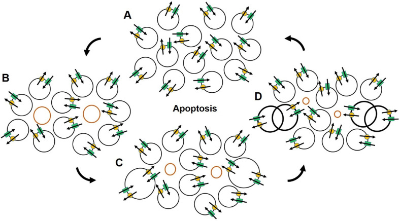
Figure 2.
The illustration depicts important characteristics of normal apoptosis that rely on proper ion channel expression and function and are essential for maintaining healthy organ structure and integrity. The circular profiles represent relative size of normal cells (black circles) and cells scheduled for programmed cell death (red circles). Parallel green bars and yellow dots represent typical ion channels and their restorative mechanisms, here for Na+, respectively. Arrows denote the direction of ionic flow. Dividing cells are indicated by overlapping circles and bold lines. Beginning in (A), normal cells for a portion of functioning organ are rendered with relatively uniform size. As apoptosis begins (B), there is a shift in ion channel function in the cells scheduled for apoptotic death (red circles) resulting in a decrease in volume that progresses through (C,D) as the process continues. This process provides room for replacement cells seen dividing in (D). Upon completion of cell division, the remains of the dead cells are removed by phagocytosis maintaining structural integrity in (A), while renewing component cells. Note that this process occurs without initiating inflammation.

