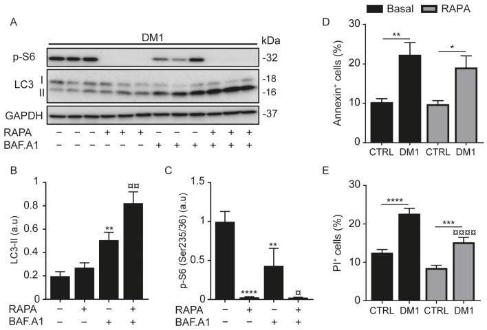
Figure 2.
Autophagy and cell death are induced in DM1 fibroblasts. (A–C) Fibroblasts from patients with DM1 were treated with 100 nM bafilomycin A1 (BAF.A1) and/or 1 μM rapamycin (RAPA) for 2 h. Protein expression levels of LC3-II (A,B) and p-S6 (Ser235/236) (A,C) were determined by immunoblotting and their densitometry was normalized to the loading control, GAPDH. Data correspond to the mean ± SD of three independent lines (** p < 0.01, **** p < 0.0001 compared with untreated cells, and ¤ p < 0.05, ¤¤ p < 0.01 versus BAF.A1-treated cells with one-way ANOVA-Tukey’s test), arbitrary units (a.u). (D,E) CTRL and DM1 cells were treated with 1 µM RAPA for 24 h and then stained with annexin and propidium iodide (PI). The percentages of annexin-positive (D) or PI-positive (E) cells were detected by flow cytometry. Data are the mean % ± SEM of three replicates (* p < 0.05, ** p < 0.01, *** p < 0.001, **** p < 0.0001 with respect to CTRL cells and ¤¤¤¤ p < 0.0001 versus DM1 cells with two-way ANOVA-Tukey’s test), N = 10,000 events. Each group (CTRL or DM1) consisted of three cell lines. All experiments were performed at least three times.

