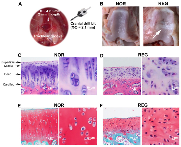
Figure 1.
The histological profiles of normal and regenerated cartilages in rabbits. (A). The microfracture (MF) surgery is conducted on the trochlear groove of the left distal femur. An articular cartilage defect (Ø ~ 4 × 6 mm, 2 mm in depth) has been created on the trochlear groove of the left distal femur with a sterilized cranial drill bit (ØO = 2.1 mm). (B). The photographs of the trochlear grooves. Compared to the normal (NOR) cartilage, the regenerated (REG) cartilage loses its transparent color and smooth and glistening appearance, replaced with a rough surface with fissures. (C,D) Examples of the H&E staining. Compared to the NOR cartilage, the REG cartilage loses the highly organized structure composed of four zones, i.e., the superficial, middle, deep, and calcified zones. (E,F) Example of the Safranin-O/Fast Green staining. Compared to the NOR cartilage, the reddish-stained ACAN content in the REG cartilage was lower. The scale bars represent 40 μm and 10 μm.

