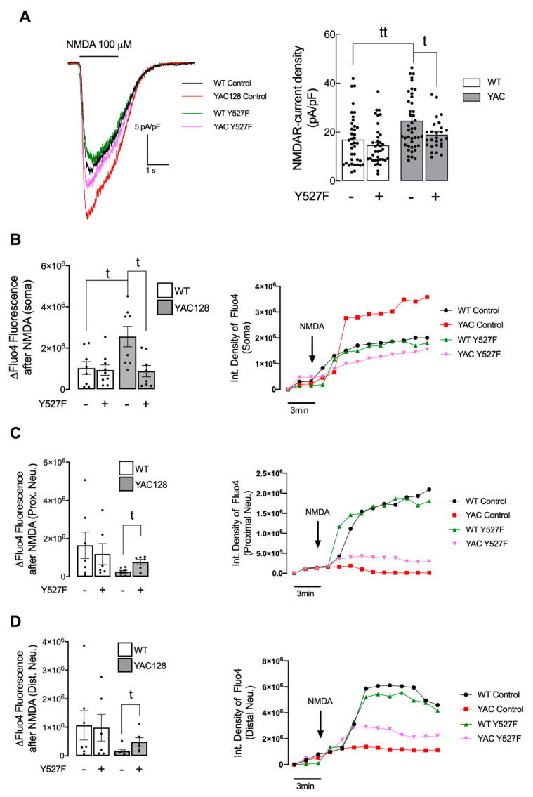
Figure 4.
Overexpression of activated SKF partially restores NMDARs currents levels in HD neurons. (A) Representative traces of NMDA (100 μM)-induced inward currents (−60 mV holding potential; with 10 μM glycine; without Mg2+) in striatal neurons from wild-type (WT) and YAC128 mice showing a higher NMDA-induced current density in YAC128 mice-derived neurons vs. WT-derived neurons, partially restored after expression of SKFY527F, as quantitatively summarized in the histogram. Data are expressed as the mean ± SEM of peak NMDA-induced current density (pA/pF). Statistical analysis: t p < 0.05, tt p < 0.01 by unpaired Student’s t-test. (B–D) Intracellular Ca2+ levels were measured using Fluo4 fluorescent dye, in soma (B), proximal neurites, (C) and distal neurites (D) units of fluorescence were monitored before (3 min) and after (15 min) exposure to 100 μM NMDA, in medium without Mg2+ and supplemented with glycine (20 μM) and serine (30 μM) in primary striatal neurons from WT and YAC128 mice, using fluorescence microscopy. Data are presented as the mean ± SEM of 4 independent experiments considering ~3 wells/condition. Statistical analysis: t p < 0.05, compared with WT control or YAC128 control (nonparametric Mann-Whitney test).

