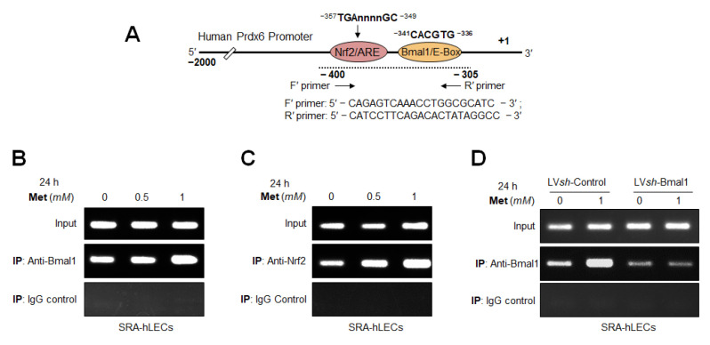
Figure 13.
In vivo DNA binding assay revealed enhanced enrichment of Bmal1 to its binding at E-Box and Nrf2 to its ARE sites of Prdx6 promoter in Metformin-treated SRA-hLECs. Diagrammatic sketch of the 5′-constructs of Prdx6 promoter containing Bmal1/E-Box- and Nrf2/ARE-DNA binding sites and ChIP primers sequences used for ChIP assay shown in Figure 12A. (B,C) DNA binding experiments with SRA-hLECs treated with Metformin was performed as noted in Figure 12. Increased binding of Bmal1 and Nrf2 to its responsive elements in the Prdx6 promoter was observed, suggesting SRA-hLECs responded similarly with Metformin as primary hLECs, suggesting SRA-hLECs are a good alternative for primary hLECs to elucidate the mechanism(s) of protective activity of therapeutic molecules. Chromatin samples were prepared from SRA-hLECs treated with different concentrations of Metformin and were submitted to ChIP assay with ChIP grade antibodies anti-Bmal1 (B) and anti-Nrf2 (C) and anti-IgG (B,C). Immunoprecipitated DNA fragments were purified and processed for PCR analysis using primers that specifically recognize fragments of Bmal1 and Nrf2 binding site (−400 to −305) of human Prdx6 gene promoter as indicated. Immunoprecipitated/pulled DNA fragments were subjected to RT-PCR analysis. Fragmented DNA was amplified with primers that specifically recognized a fragment of human Prdx6 promoter containing E-Box or ARE site. 10% sheared chromatin was used as input. Control IgG was used as negative control. (D) Bmal1 knockdown SRA-hLECs involvement of Metformin to enhance the Bmal1 binding to E-Box in the Prdx6 promoter in vivo. SRA-hLECs were stably infected either with GFP (green fluorescence protein) linked lentiviral (LV) Sh-control or GFP-linked LV Sh Bmal1 as described materials and Methods section. Genomic DNA isolated from LV Shcontrol and LV ShBmal1 SRA-hLECs were cross-linked to immobilize bound protein in vivo, was sheared and immunoprecipitated with anti-Bmal1 or control IgG. Immunoprecipitated DNA was amplified using PCR with specific primers. 10% chromatin was used as input DNA. Amplified DNA band visualized with ethidium bromide staining as shown in photographic images.

