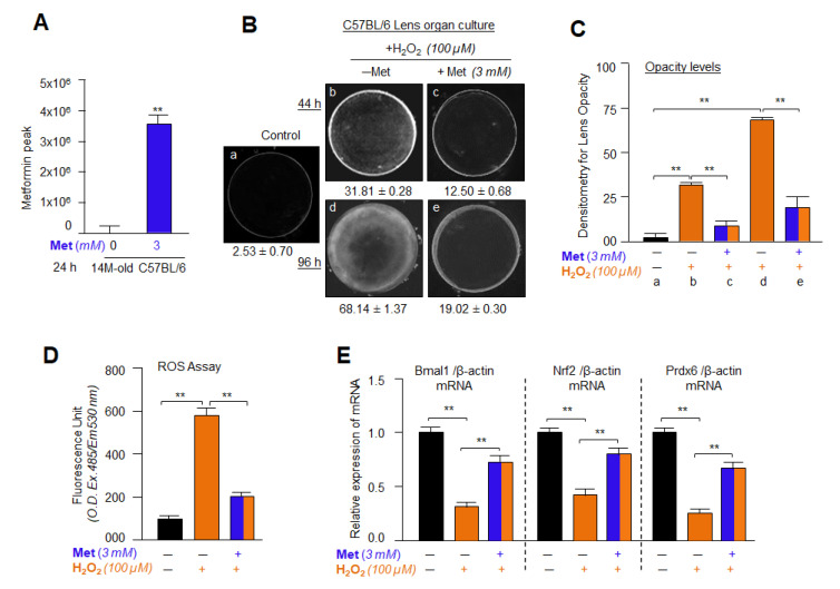
Figure 16.
Metformin successfully internalized into mouse eye lenses and delayed/prevented the lens opacity and mitigated ROS accumulation induced by H2O2 by enhancing the Bmal1/Nrf2/antioxidant, such as Prdx6 in vitro. (A) Metformin penetration in the mouse lens cultured in vitro. Lenses isolated from 14 months old mouse were cultured in 48-well culture plate and treated with 3 mM Metformin for 24 h and examined for Metformin penetration using Acquity UPLC coupled with Waters triple-quad Xevo TQS analysis and Metformin peak was presented as histogram. The data represent the mean ± S.D. from three independent experiments. Untreated vs. Metformin-treated; ** p < 0.001. (B) Lenses isolated from 14 months old C57BL/6 mouse were used for in vitro organ culture as described previously [67,68,85]. The cultured lenses were treated with 3 mM of Metformin followed by 100 µM of H2O2 exposure. 44 h or 96 h later the lenses were photographed (Nikon SMZ 745T) connected with Camera and software (Nikon). Photograph is representative of three experiments: Metformin significantly prevented the H2O2-induced lens opacity by 61% and 72% (c and e) compared to H2O2 (b and d) and untreated lens control (a). (C) Histogram indicates densitometry of lens opacity. a, 2.53 ± 0.70 (control, black bar); b, 31.81 ± 0.28 (orange bar: H2O2 alone, 44 h); c, 12.50 ± 0.68 (blue-orange bar: Metformin + H2O2 alone, 44 h); d, 68.14 ± 1.37 (orange bar: H2O2, 96 h); e, 19.02 ± 0.30 (blue-orange bar: Metformin + H2O2, 96 h). All histograms are presented as mean ± S.D. values-derived from three independent experiments, ** p < 0.001. (D,E) Lenses Isolated from 14 months old C57BL/6 mouse were cultured and treated with 3 mM of Metformin followed by H2O2 exposure. 48 h later ROS level and mRNA expression of Bmal1, Nrf2 and Prdx6 were analyzed using H2-DCF-DA dye and RT-qPCR analyses, respectively as indicated in Figure, and described in Materials and Methods. All histograms are presented as mean ± S.D. values-derived from three independent experiments, ** p < 0.001.

