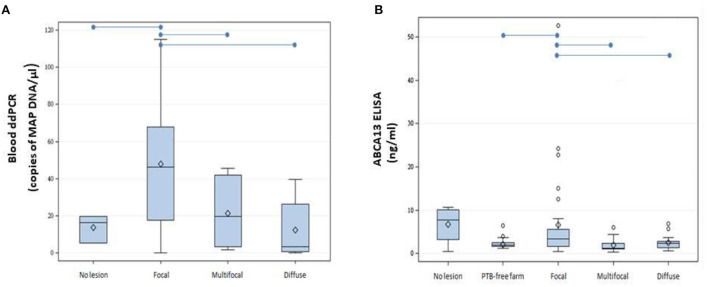
Figure 2.
Boxplots showing the distribution of ABCA13 levels and copies of MAP DNA/μl obtained using blood ddPCR. (A) Distribution of copies of MAP DNA/μl obtained using blood ddPCR in cows without lesions and with different PTB-associated lesions. (B) Results of the ABCA13 levels measured by ELISA in serum samples of 61 cows from the PTB-free farm and from 74 slaughtered cows without lesions (N = 4) or with focal (N = 32), multifocal (N = 21), and diffuse lesions (N = 17) in gut tissues and lymph nodes. The plots represent the interquartile range and the whiskers represent the 95% range. Lines and diamonds within the boxes represent the median and the mean of each group, respectively. Blank dots represent outliers. Blue lines on the top of the figures represent statistically significant differences (P ≤ 0.05).

