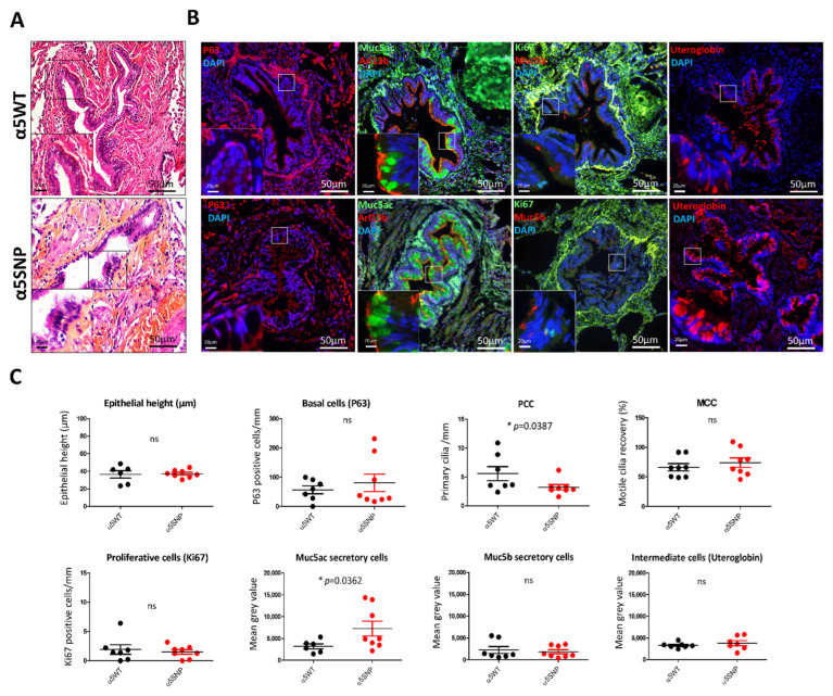
Figure 2.
Bronchiolar epithelial remodeling in rs16969968 (α5SNP) COPD patients. (A): Hematoxylin and eosin staining showing the epithelial height of α5SNP and α5WT COPD patients. (B): Examples of the microscopic acquisition of immunofluorescent stainings for basal cells (P63, red), ciliated cells (Arl13b, red), proliferative cells (Ki67, green), mucins secretory cells (Muc5ac, green; Muc5b, red), and intermediate cells (Uteroglobin, red). Nuclei are stained in blue (DAPI). Magnification corresponding to the selected area is represented. (C): Dot plots (means with SEM) representing measurements of the epithelial height, the number of basal, proliferative, and primary ciliated cells per mm, motile cilia recovery (%), and the mean grey values of mucins (Muc5ac, Muc5b) and uteroglobin-associated fluorescence of α5SNP and α5WT COPD patients. *, p < 0.05 α5WT vs. α5SNP; ns, non-significant.

