Figure 3.
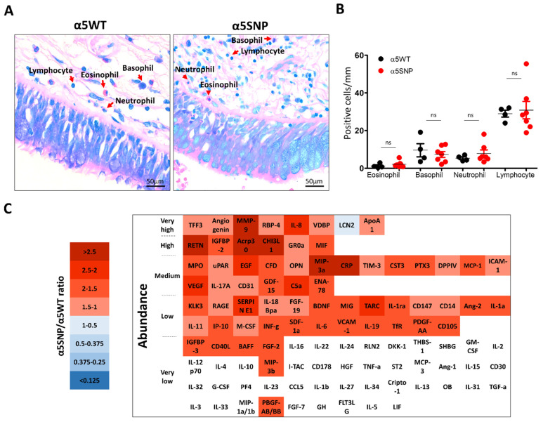
Lung inflammatory response in rs16969968 (α5SNP) COPD patients. (A): Microscopic acquisitions showing peribronchial recruitment of immune populations in α5SNP and α5WT COPD patients. (B): Dot plot showing the number of eosinophils, basophils, neutrophils, and lymphocytes per mm of epithelium in α5SNP vs. α5WT COPD patients. (C): Heatmap presenting the ratios of inflammatory mediators’ expression in broncho-alveolar lavage fluids of α5SNP vs. α5WT COPD patients. Downregulated inflammatory mediators are presented in blue, and upregulated ones are in red. The inflammatory mediators whose expression is lower than the detection cut-off value (5% of positive control) are identified in white. The inflammatory mediators are categorized according to their detected abundance in the broncho-alveolar lavage fluids of COPD patients (from very high, >50% of the detection of the positive control; to very low, <5% of the detection of the positive control). ns, non-significant.

