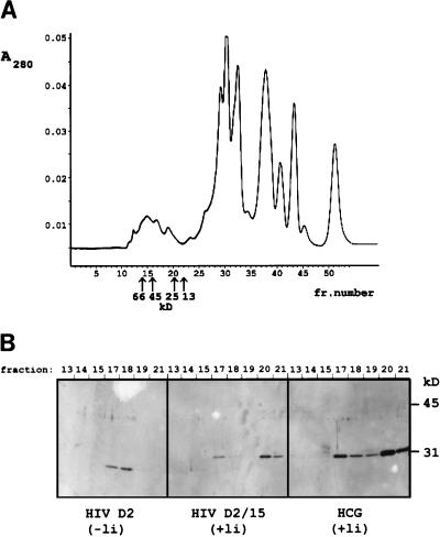
FIG. 3.
Gel filtration of periplasmic fractions on Superdex 75HR and analysis of the collected fractions on a Western blot with anti-FLAG M2. (A) Chromatogram, obtained by plotting A280 against the fraction number, illustrating the separation of the periplasmic proteins from clone D2. The retention times of the marker proteins are indicated with arrows. (B) Western blot analysis (15% gel) of the collected fractions (fraction number is shown above the blots) obtained from gel filtration of anti-p24 clones and of an anti-hCG scFv-producing clone. Two peak fractions can be discriminated in the scFvs having a complete linker (the derivative D2/15 and the anti-hCG): a main peak of the monomer (fraction 20; molecular mass, 25 kDa) and a dimer peak (fraction 17; molecular mass, 45 kDa). scFv of clone D2 emerged from the column in a single peak corresponding to the dimeric product.

