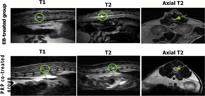
Fig. 5.
MRI analysis of the spinal cord. The EB-treated group is characterized by a large hypointense lesion (circle) on sagittal T1 scan and a diffuse hyperintense lesion on sagittal (circle) and axial (arrow) T2 scans while the PRP co-treated group has a small faint hypointense lesion on sagittal T1 scan and a faint hyperintense lesion on sagittal and axial T2 scans showing decreased intensity and extent of the lesion (circle and arrow)

