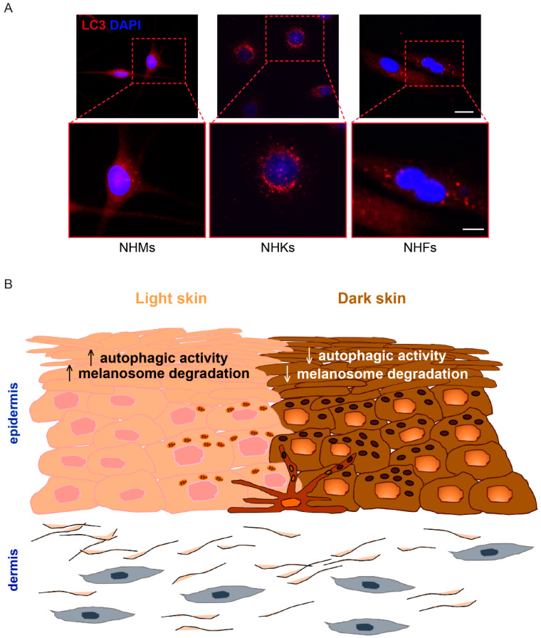
Figure 1.
Skin color and autophagy. (A) Immunofluorescence analysis was performed on primary cultures of normal human melanocytes (NHMs), normal human keratinocytes (NHKs) and normal human fibroblasts (NHFs) using an antibody (red signal) directed against the autophagy marker light chain 3 (LC3I/II). Nuclei are counterstained with DAPI (blue signal). Scale bars: 20 μm; enlarged view of the boxed area: 10 μm. (B) Autophagic activity is involved in skin color variation by regulating melanosome degradation.

